muscle fiber orientation
Ca ions are pumped back into the SR. Fiber orientations were determined by measuring fiber angle relative to the circumferential direction helix angle.

Marginal M Posterior P And Anterior A Fibers In The Soleus Download Scientific Diagram
Analysis the orientation of muscle fibers in planarians.

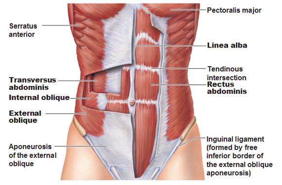
. This study aimed to measure muscle fibre orientation and other parameters of muscle morphology of the abdominal muscles in relation to palpable bony landmarks and found that the fibres of obliquus externus abdominis were about 4 degrees more vertical than the lower edge of the eighth rib. Click again to see term. The muscle fibres of upper rectus abdominis were 2 degrees inferolateral to the midline while the lower rectus abdominis muscle fibres deviated inferomedially from the midline by about 8 degrees.
Skeletal muscle fiber orientation is a critical factor for the success of this procedure. The metabolite profiles were different for each orientation of muscle fibers to the main magnetic field. The same terminology is used when referring to the relative alignment of the noncontractile tissues for example tendons and epimysium.
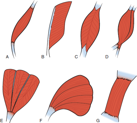
In the inner layer the inclination was. The dense muscle fiber microstructure gives rise to orientation dependent MR features with anisotropic overall motion of the creatine Cr and phosphocreatine PCr molecules causing residual dipolar couplings first described for the total observed creatine tCr CrPCr resonances 1 2 while orientation dependence was later also reported for. Parallel strap have fibres which as the name suggests run parallel to each other.
Analysis the orientation of muscle fibers in planarians - GitHub - walkernoreenmuscle_fiber_orientation. Figure 1033 Contraction of a Muscle Fiber. Fibers are running perpendicular or at right anles with the midline or.
Relaxation of a Muscle Fiber. There was no variation of muscle fiber orientation in the ischiatic head of BF from the proximal to the distal. Orientation of muscle fibers was determined.
Muscle fibers of the ischiatic head of BF were parallel to the long axis of the muscle. Tap card to see definition. Smoothly varying muscle fiber orientations in the heart are critical to its electrical and mechanical function.
Our results indicated that skeletal muscle fiber oriented circumferential to the heart and perpendicular to the ventricular septum is the preferred procedure for dynamic cardiomyoplasty. Click card to see definition. 2294346 Indexed for MEDLINE Publication Types.
The traditional manual method for MFO estimation in sonograms was labor-intensive. However this is an indirect. There are many possible ways to specify the complex fibre orientations in a finite element model for example defining a local element coordinate system.
The orientation of fibers influences the overall function of the muscle Richards 2008. The automatic methods proposed in recent years also involved voting procedures which were computationally expensive. Alternatively fast-twitch IIa and IIx fibers are abundant in elite power.
Tap again to see term. Muscle fibers that align end to end with each other are referred to as being in series while fibers that align side by side are referred to as parallel fibers. The fraction of the myocardial volume occupied by connective tissue was determined by point counting.
Differences less than 005 were considered to be statistically significant with 95CI. As long as Ca ions remain in the sarcoplasm to bind to troponin and as long as ATP is available the muscle fiber will continue to shorten. Human muscle fibers are generally classified by myosin heavy chain MHC isoforms characterized by slow to fast contractile speeds.
Fibers run straight or parallel with the midline or longtidunal axis of the body or limb. In addition the other fifteen sides will be cut into boneless subprimal cuts and subsequently into 25-cm thick steaks that would be typical for each cut. Skeletal muscle tissues have complex geometries.
This fiber orientation provides more range of motion but less power for contraction. In addition the complex fibre orientation arrangement makes it quite difficult to create an accurate finite element muscle model. A cross-bridge forms between actin and the myosin heads triggering contraction.
Some textbooks include Fusiform muscles in the parallel group. The ability of surface electrodes to accurately detect the activity of a particular. The orientation at 90 was the most.
Measurements using common surface landmarks were used to determine the relationship of these muscles with the landmarks eg biceps muscle bulk extends from the upper fourth to the lower fourth of the humerus. They are normally long muscles which cause large movements are not very strong but have good endurance. A muscle fiber orientation in which the component skeletal muscle cell is an equal distance apart for the entire length of the fibers everywhere.
Muscle fiber orientation in the left ventricular myocardial layer was histometrically estimated in normal concentric and eccentric hypertrophied hearts. The muscle fiber orientation will be determined for each slice such that a three-dimensional map of the muscle fiber orientation for each muscle can be created. Inclination of muscle fibers of the long head BF were gradually more angular to the horizontal axis of the muscle.
The appropriate surface electrode placements which follows the muscle fibre orientation of the obliquus externus abdominis obliquus internus abdominis and rectus. Muscle fiber orientation MFO is an important parameter related to musculoskeletal functions. From detailed histological studies and diffusion tensor imaging we now know that fiber orientations in humans vary gradually from approximately 70.
6 rows Muscle fiber orientation can be determined from dipolar coupling. The angle of inclination of muscle fibers from coronal section was largest in the innermost and outermost zones and was progressively diminished toward the middle layer in all the hearts. Metabolites of interest from each orientation to the main magnetic field were compared using Wilcoxon signed-rank test.
Type I or slow-twitch fibers are seen in high abundance in elite endurance athletes such as long-distance runners and cyclists. There was significant P 0001. Start studying Orientation of Muscle Fibers.
Examples include Sartorius and Sternocleidomastoid. Learn vocabulary terms and more with flashcards games and other study tools.

Fascicle An Overview Sciencedirect Topics

Muscle Fiber An Overview Sciencedirect Topics

11 2 Explain The Organization Of Muscle Fascicles And Their Role In Generating Force Anatomy Physiology

Pennate Muscle An Overview Sciencedirect Topics
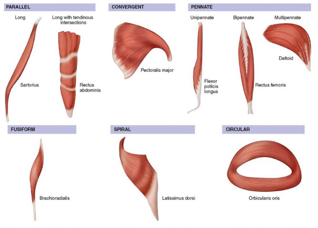
How Muscles Work Part 2 Of 2 Shapelog

Pennate Muscle An Overview Sciencedirect Topics
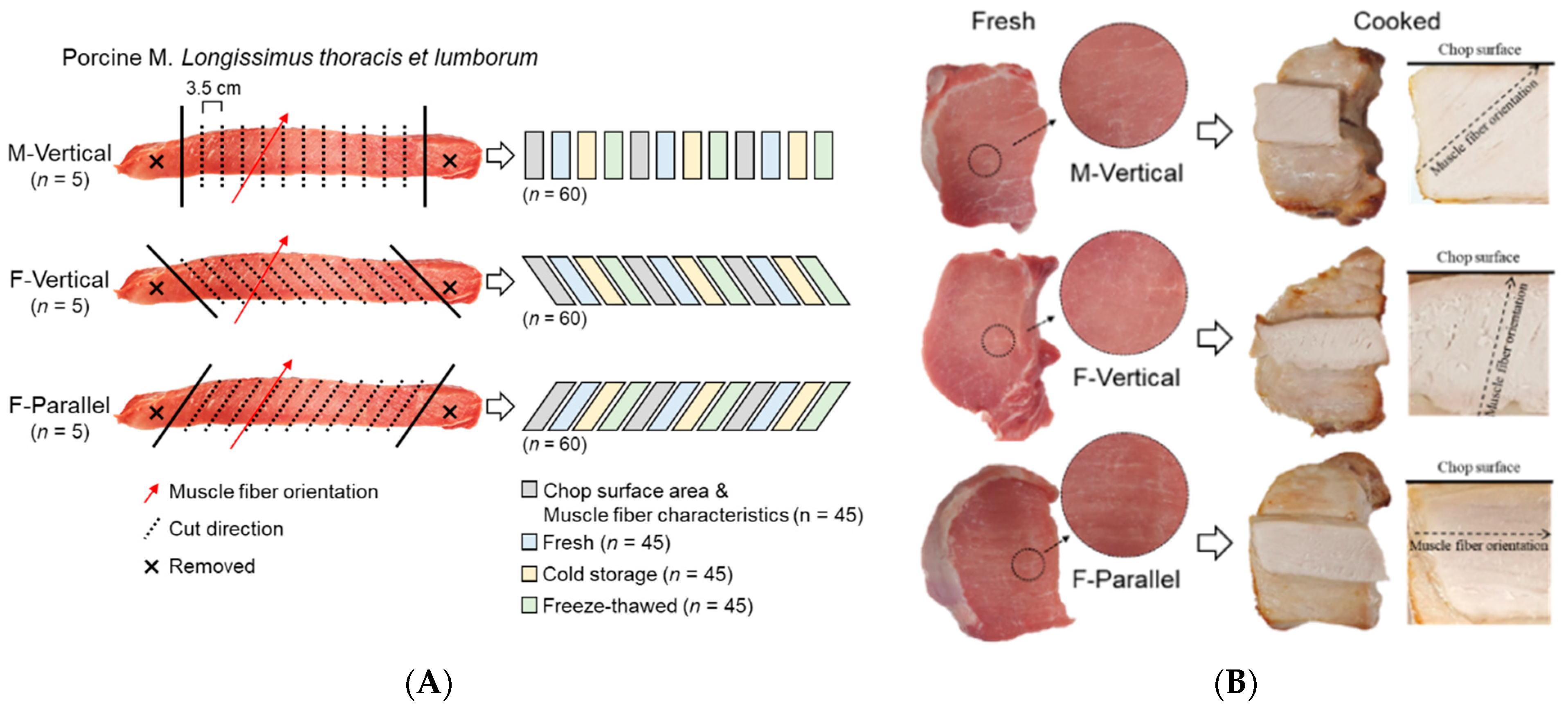
Foods Free Full Text Pork Loin Chop Quality And Muscle Fiber Characteristics As Affected By The Direction Of Cut Html
Organization Of Skeletal Muscles Course Hero

The Heart Location Of Heart Surrounded By Pericardium 1 5cm Left From Center Size Of A Fist G 흉골 胸骨 Ipad Mini 308g 심낭 심막 Ppt Download

Classification Of Muscles Based On The Directions Of Muscle Fibers The Download Scientific Diagram
Organization Of Skeletal Muscles Course Hero

3 Orientation Of Cardiac Muscle Fibers Download Scientific Diagram
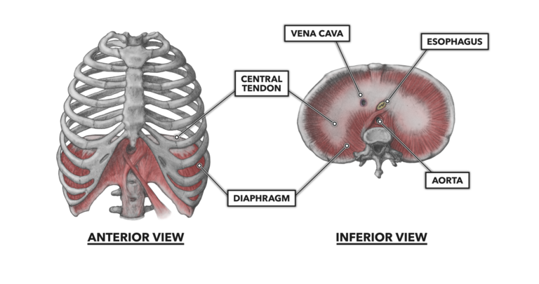
Crossfit Thoracic Muscles Part 2
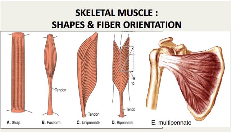
Muscle Histology Flashcards Chegg Com

Fdi Muscle Fiber Tracks Derived From The Dwi Data In Three States At Download Scientific Diagram
Comments
Post a Comment